


by László R. Orzó, Zoltán Á. Varecza, Márton Zs. Kiss and Ákos Zarándy (MTA SZTAKI)
Digital holographic microscopy (DHM) provides a simple way to automate biological water monitoring. Using proper computational imaging algorithms, the rather complicated measuring techniques can be replaced by easy methods, and the further data processing steps are also simplified.
Companies that utilise large amounts of water (e.g., water-works, fisheries, etc.) regularly need information about its microbiological content. These microbiological data can be regarded as one of the quality indicators of the water.
The conventional microbiological measurement techniques require sampling the water and completing some complex preprocessing steps. The organisms within the samples must be identified and classified by a skilled human observer using an appropriate microscope setup. This process is extremely difficult to automate.
However, digital holographic microscope technology makes automation much easier. One of the main advantages of digital holographic microscopy is that it can analyse much larger volumes than conventional microscopes, where the small depth of field limits the observable volume considerably. The device using holographic technology does not record sharp, focused images, but detects all the objects in the volume measured. Based on the digitally measured hologram, the images of all objects in the volume observed can be reconstructed by using proper numerical methods. This task can be fulfilled even if the objects are far from the actual focal plane of the microscope. Consequently, the small depth of field limitation of the conventional microscopy can be eliminated, and 200 times larger volumes can be analysed correctly by this method. Hence, the otherwise indispensable sample condensation step can be omitted completely. In the digital holographic microscope the sample is moved by a precision pump and measured in the flow-cell. The scheme of the DHM measuring setup is shown in Figure 1.
Figure 1: The scheme of the DHM measuring setup.
Detection and segmentation of the objects measured in a flow-cell is also a simple task. The image of the objects in such a cell can be easily segmented utilising their 3D position information within the volume, while in a settled sample they frequently overlap each other. Using a proper algorithm, the holograms of the reconstructed objects can be removed from the original hologram and this way the diffractions of the objects nearby will not interfere with the imaging of the objects of interest [1]. An appropriate classification algorithm is implemented into the system as well based on the use of the reconstructed images of the objects. Implementing all the technologies described above, a complete, automatic, water monitoring system has been constructed. Using stream processors massively parallel computing capacities, the hologram reconstruction process is considerably accelerated, and close to real time processing has been achieved. The scheme of the DHM based water monitoring system is shown in Figure 2.
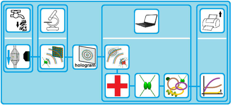
Figure 2: The scheme of the Digital Holographic Microscope based water monitoring system. It includes: the recording of the color holograms of the objects within the sample using Digital Holographic Microscope, the appropriate numerical reconstruction of the image of the objects, with the compensation of some of the measuring setup aberrations, and finally, the morphology, size and color based classification of the different microorganisms.
In our projects we have developed three different DHM devices.
The first device detects algae and other microorganisms. It is able to measure the images of objects with 1µm lateral resolution in the flow cell. The observed volume of a single hologram is 1µl. Since colour information is usually indispensable for the correct classification of the different algae, we record three different colour holograms. We apply red, green and blue fibre coupled lasers and record the different colour holograms simultaneously using a colour digital area scan sensor. The crosstalk between the different coloured pixels is numerically compensated. The fibre ends do not need to be in exactly the same position since using proper numerical methods it can be corrected [2,3]. The use of the colour, shape and size information of the reconstructed algae images results in an appropriate object classification. Continuous monitoring can correctly outline the distribution and numerical changes of the different alga species within the consecutive samples.
The second device images and monitors larger creatures (e.g., nematodes, rotifers). This device has only 3µm lateral resolution but the observable volume is 500µl. This device is able to measure up to 1 litre of fluid sample within 20 minutes and so the temporal change of the number of worms can be continuously examined. It can be used to monitor filter quality in waterworks wells since an increasing number of worms can indicate contamination of the filters, meaning that cleaning is required. Such measurements can also be applied to check the efficiency of the cleaning process.
The third device combines fluorescent detection with volumetric imaging of the digital holographic microscopy. It detects the position of the (auto-) fluorescent objects and reconstructs a high resolution image of these objects from a simultaneously measured hologram. Using this device, by the application of a proper fluorescent dye (e.g., fluorescein diacetate) the separation of live and dead objects is also achievable within a large volume, while their high resolution images are also reconstructed. This could also be used to measure the efficiency of a ballast water treatment procedure (NIVA – Norwegian Institute for Water Research).
The commercial release of the devices described above is in progress (WaterScope International Inc.).
The microbial community structure in the microbiome of a lake, ballast-water or drinking water shows location and time dependency. The more accurate and more prompt a monitoring method is, the more efficient the interventions can be. The risk of appearance of toxic alga species in sea water is a continuous threat not only for sea fish-farms but also for coastguards maintaining the quality of water on the shoreline. The series of instruments designed and developed at the Hungarian Academy of Sciences combine state-of-the-art imaging technologies with advanced computation algorithms and accurate microbial classification system to offer a new generation of water monitoring instruments. The multi-purpose use of these instruments offers the possibility for different industries and government agencies to maintain water quality at a much lower cost and - by the early warning function - eliminate risks based on certain species of microorganisms detected.
Our computational imaging methods offer a far simpler solution to biological water monitoring than conventional techniques.
This projects were funded by the Hungarian National Office for Research and Technology (NKTH 1981822A) and the Framework of Green Industry Innovation Programme of the EEA and Norwegian Financial Mechanisms (grant number: HU09-0078-A12013).
Links:
http://www.analogic.sztaki.hu
http://waterscope.eu/en/
References:
[1] L. Orzó, Z. Göröcs, A. Fehér, and S. Tőkés: “In-line hologram segmentation for volumetric samples. Applied Optics”, 52(1): A45–A55, (2013).
[2] M. Zs. Kiss, et al.: “Special multicolor illumination and numerical tilt correction in volumetric digital holographic microscopy”, Opt. Express 22, 7559-7573 (2014).
[3] L. Orzó: “High speed phase retrieval of in-line holograms by the assistance of corresponding off-axis holograms”, Opt. Express 23(13), 16638-16649 (2015).
Please contact:
László R. Orzó, MTA SZTAKI, Budapest Hungary
+ 36 06 1 2797182

