


by Samuli Siltanen (University of Helsinki)
X-ray tomography is a wonderful tool that allows doctors to peek inside patients. However, since X-rays are harmful, a patient’s exposure to them should be limited. An obvious way to achieve this is to take fewer images, but unfortunately this causes trouble for classical image reconstruction methods. However, new mathematical methods, based on compressed sensing and the multi-scale shearlet transform, can save the day!
The goal of medical tomography, or CT scan, is to reveal the inner structure of a patient using a collection of X-ray images recorded from several directions. In a classical setup, 360 images are taken while rotating around the patient with 1 degree angular steps, and a filtered back-projection (FBP) is used to create a highly detailed reconstruction image. My research team at University of Helsinki aims at reducing the number of images and using shearlet sparsity for reconstruction.
Imaging the patient from hundreds of directions exposes him or her to a substantial radiation dose. Therefore, such comprehensive CT scans are limited to rather serious medical conditions so that the dose is ethically acceptable.
Within the medical community there is a growing interest in taking fewer X-ray images and using them to compute a less-than-perfect tomographic reconstruction that is good enough for a particular purpose. For example, in dental implant planning it is enough to know the depth and direction of the desired hole to be drilled. Likewise, in surgery it may be enough to get an idea of the 3D position of instruments during a knee operation. In both of these cases, there exists a device (panoramic X-ray imager and surgical C-arm device, respectively) for collecting images from a range of directions.
The mathematical task of reconstructing the tomographic image becomes more difficult when there are fewer images. The measurement information alone is not sufficient to perfectly determine the 3D target structure, and the inversion is extremely sensitive to noise and modelling errors. These problems can be overcome by complementing the insufficient data with a priori knowledge about the target, in other words by using regularisation.
One way to regularise the tomographic reconstruction problem is to promote sparsity in the spirit of [1,2]. This means expressing the unknown image in terms of suitable building blocks and requiring that as few building blocks as possible are used. Traditional choices for building blocks include sines and cosines arising from fast fourier transform (FFT) and multiscale constructions such as wavelets. Both of them have serious shortcomings in the context of tomography, however. FFT is not good for local details as sines and cosines span the whole image area. Wavelets in turn, make it possible to zoom in to details, but they are not economical for representing jumps in X-ray attenuation along tissue boundaries, the very things doctors most often want to see.
The shearlet transform [3] is optimal for representing jumps along curves in images. The optimality is achieved by making the finer-scale building blocks more and more elongated as they become smaller. Additionally, the smaller these needle-like image atoms are, the more possible orientations they have. Thus, shearlets allow faithful following of tissue boundaries.
Figure 1 shows three reconstructions of a walnut. (The data is openly available for experimentation, see [L1]. On the left is reconstruction from comprehensive data with 1200 X-ray images. Note the high level of detail delivered by the standard FBP algorithm. The middle and right images are reconstructions computed from the same subset of only 20 X-ray images taken all around the walnut. The middle image shows FBP reconstruction; we remark that FBP was never designed for this kind of data with large angular steps between projection directions. Shown on the right is reconstruction based on shearlet sparsity.
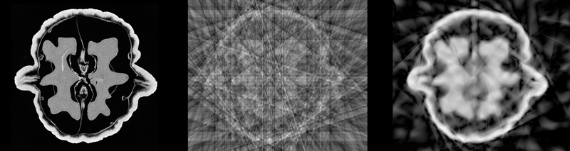
Figure 1: Three reconstructions of a walnut.
The high quality of the sparse-data walnut reconstruction inspired my team to collaborate with Professors Miika Nieminen and Simo Saarakkala at Oulu University Hospital, Finland. Together we aim to take shearlet-sparsity regularisation to clinical practice, speeding up radiological examinations and reducing harmful radiation doses to patients.
Link:
[L1] http://fips.fi/dataset.php
References:
[1] I. Daubechies, M. Defrise, and C. De Mol: “An iterative thresholding algorithm for linear
inverse problems with a sparsity constraint”, Communications on pure and applied mathematics, 57, 2004.
[2] K. Hamalainen, et al.: “Sparse tomography”, SIAM Journal of Scientific Computing 35, 2013, DOI:10.1137/120876277
[3] G. Kutyniok, D. Labate: “Shearlets: Multiscale Analysis for Multivariate Data”, Birkhäuser, 2012.
Please contact:
Samuli Siltanen
University of Helsinki
+358 40 594 3560

